Sunday, October 25, 2009

DENS EVAGINATUS

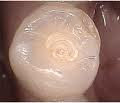
Dens envaginatus is a condition found in teeth where the outer surface appears to form an extra bump or cusp. Premolars are more likely to be affected than any other tooth. This may be seen more frequently in Asians. The pulp of the tooth may extend into the dens evaginatus. There is a risk of the dens evaginatus chipping off in normal function. Hence this condition requires monitoring as the tooth can loose its blood and nerve supply as a result and may need root canal treatmentDens evaginatus (DE) is an uncommon dental anomaly, having been well documented since 1925.

It occurs primarily in people of Asian descent and is exhibited by protrusion of a tubercle from occlusal surfaces of posterior teeth, and lingual surfaces of anterior teeth.

Tubercles have an enamel layer covering a dentin core containing a thin extension of pulp. These cusp-like protrusions are susceptible to pulp exposure from wear or fracture because of malocclusion, leading to pulpal complications soon after eruption.

review of the literature reveals an abundance of terms for this anomalous dental structure, the most frequently encountered being: odontome, odontoma (odontome) of the axial core type, evaginatus odontoma (evaginated odontome), occlusal enamel pearl, occlusal tubercle, tuberculum anomalous, accessory cusp, supernumerary cusp, interstitial cusp, tuberculated cusp, tuberculated premolar, Leong’s premolar, and talon cusp (specifically for anterior teeth)

Talon cusp originated as a descriptive term for DE when observed on the lingual surface of anterior teeth because of a resemblance to an eagle’s talon. Currently, DE is the preferred terminology utilized to describe this developmental abnormality, first recommended by Oehlers in 1967


DE is thought to develop from an abnormal proliferation and folding of a portion of the inner enamel epithelium and subjacent ectomesenchymal cells of the dental papilla into the stellate reticulum of the enamel organ during the bell stage of tooth formation 

TALONS CUSP


talon cusp, also known as an "eagle's talon", is an extra cusp on an anterior tooth. The term refers to the same condition as dens evaginatus, but the talon cusp is the manifestation of dens evaginatus on anterior teeth. The incidence has been found to range from less than 1% to 6% of the population.[1] Of all cases, 55% occur on the permanent maxillary lateral incisor, and 33% occur on the permanent maxillary central incisor. They are found rarely in primary teeth.

Taurodontism


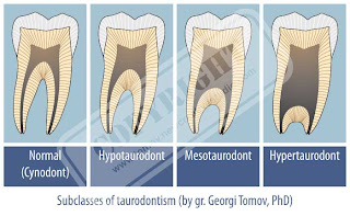
The term taurodontism was first introduced by Arthur Keith in 1913. It refers to a tooth form characterized by an external block configuration with an elongated body tending to enlarge at the expense of the root.The taurodontic teeth are identified by elongated pulp chambers and apical displacement of bifurcation or trifurcation of the roots. Due to this, the chamber has a greater apicoocclusal height than normally and lacks the constriction at the level of the CEJ giving it a rectangular shape. Etiology of taurodontism is diverse commonly attributed to the failure of invagination of the epithelial root sheath sufficiently early to form the cynodont. Autosomal transmission of the trait has also been observed. Taurodontism can occur alone limited to one or more teeth or it can be associated with various syndromes. Taurodontism may be unilateral or bilateral and affects permanent teeth more frequently than primary teeth. Taurodontism may be classified as mild, moderate and severe (Hypo, Meso and Hyper respectively) based on the degree of apical displacement of the pulpal floorTaurodontism is a condition found in teeth where the body of the tooth and pulp chamber is enlarged. As a result, the floor of the pulp and the furcation of the tooth is moved apically down the root. The term means "bull like" teeth derived from similarity of these teeth to those of ungulate or cud chewing animals.
According to Shaw these can be classified as hypotaurodont, hypertaurodont and mesotaurodont.
According to Mangion taurodontism may be:
1. A retrograde character
2. A primitive pattern
3. Mendalian recessive character
4. Atavistic feature
5. A mutation
The condition is of anthropological importance as it was seen in Neanderthals. It has also been reported in Klinefelter's syndrome. The teeth involved are invariably molars, sometimes single and at the other times multiple teeth may be involved. The teeth themselves may look normal and do not have any particular anatomical character on clinical examination.
On dental radiograph, the involved tooth looks rectangular in shape without apical taper. The pulp chamber is extremely large and the furcations may be only a few millimeters long at times.

Saturday, October 24, 2009